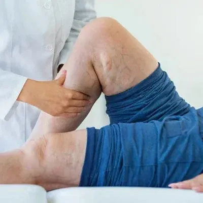

If you have varicose veins, swollen legs or aching that worsens through the day, the most useful test is a venous Doppler scan, also called a venous duplex ultrasound. It is painless, uses no radiation, and shows the information that decides your treatment.
At a sonography and vascular clinic, this is usually the first thing we do, because what you see on the skin is rarely the full story.
What is a venous Doppler scan?
It combines two kinds of ultrasound:
- Ultrasound images (B-mode) show the veins, their size and walls, and whether there is any clot inside.
- Doppler shows the direction and speed of blood flow, so we can see whether blood moves up towards the heart, as it should, or falls back down the leg because of faulty valves (reflux).
Together, this is called a duplex scan.
What the scan shows
1. Which veins are leaking, and where
The scan traces the main surface veins (the great saphenous vein on the inner leg and the small saphenous vein at the back of the calf), the deep veins, and the “perforator” veins that connect them. It shows exactly where reflux starts and how far it travels.
2. How big and deep the veins are
Vein diameter, depth under the skin and how twisted the vein is help decide between laser ablation, foam sclerotherapy or other options. Compare treatment options.
3. Whether the deep veins are healthy
Before closing a surface vein, we need to know the deep veins can carry blood out of the leg. The scan checks for blockage or damage from previous clots.
4. Blood clots (DVT)
A venous Doppler is the standard first test when deep vein thrombosis is suspected, for example sudden swelling of one leg with calf pain. Learn about DVT.
5. Why veins came back after earlier treatment
In recurrent varicose veins, the scan shows the new source of reflux and the vein “map” after previous procedures. Why varicose veins come back.
Why treatment should never start without it
Visible bulging veins are usually fed by a deeper leaking vein that can’t be seen or felt. Treating only the visible veins, without the scan, is one of the most common reasons varicose veins return within months. International guidelines recommend duplex ultrasound for everyone being assessed for varicose vein treatment.
How the scan is done
- You change into shorts or a gown so the legs are uncovered from groin to ankle.
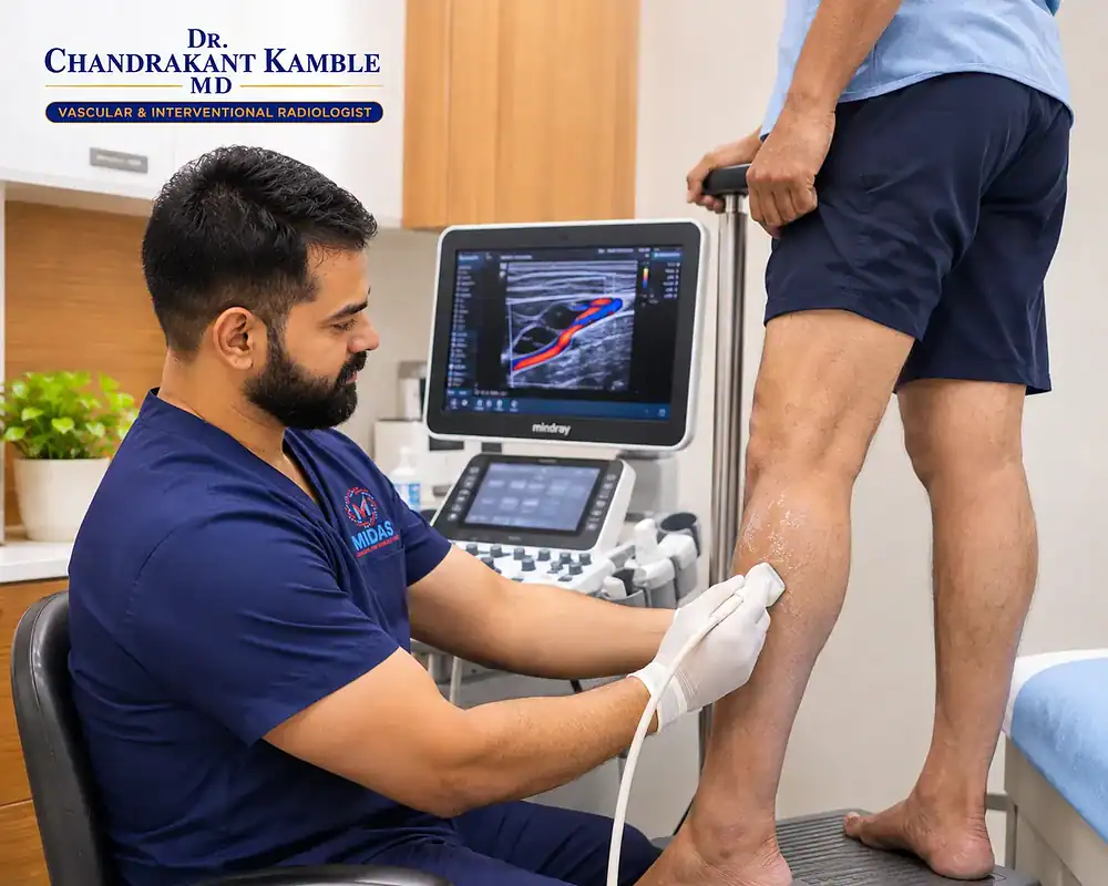
- Much of the scan is done standing, or lying with the legs lowered. Reflux is best seen when gravity pulls blood down the leg.
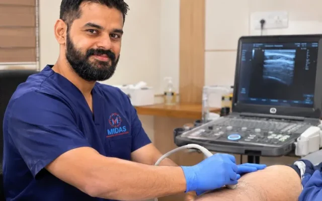
- Gel is applied, and a handheld probe is moved along the leg.
- To test the valves, the sonographer or doctor may squeeze your calf briefly or ask you to bear down, as if blowing into a closed fist. If the valve is faulty, the Doppler shows blood falling back for longer than normal.
- Both legs are usually scanned, even if only one has symptoms.
It is painless. You may feel mild pressure from the probe and calf squeezes. A full study of both legs commonly takes 30–60 minutes.
How to prepare
- No fasting is needed; eat and drink normally.
- Wear loose, comfortable clothing.
- Don’t apply creams or oils to the legs that day.
- If you wear compression stockings, you can wear them to the appointment; they’ll be removed for the scan.
- Bring previous scan reports, procedure details and a list of medicines, especially blood thinners.
Understanding your report
Your report will mention:
- the veins examined and whether each has reflux, usually with timing. In the surface veins, reflux lasting longer than about half a second is considered abnormal.
- vein diameters, in millimetres
- the presence or absence of thrombus (clot)
- the perforator veins involved
Terms like “incompetent GSV” or “SFJ reflux” mean the great saphenous vein, or its junction with the deep vein in the groin, is leaking. Your doctor will explain what it means for you and draw your personal vein map.
Scan and plan at one visit in Vasai
At Midas Sonography & Vascular Clinic, Ambadi Road, Vasai (W), the venous Doppler is performed and interpreted by Dr. Chandrakant S. Kamble, a vascular & interventional radiologist. The doctor who reads the scan is the same specialist who plans and performs your treatment, so you leave the first visit knowing exactly what is wrong and what can be done.
The clinic is open Monday to Saturday, 8 AM – 9 PM, convenient for patients from Vasai, Virar, Nalasopara, Naigaon, Mira-Bhayandar, Palghar and Boisar.
About varicose veins treatment · Book a scan and consultation
Frequently asked questions
Is a venous Doppler scan painful?
No. It uses gel and a handheld probe. You may feel mild pressure and brief calf squeezes.
Is there any radiation?
No. Doppler ultrasound uses sound waves and is safe, including in pregnancy.
Do I need to fast before a leg Doppler?
No. You can eat and drink normally. Wear loose clothing.
What is the difference between a venous and an arterial Doppler?
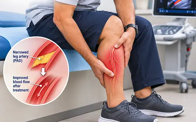
A venous Doppler checks the veins that return blood to the heart, for varicose veins and clots. An arterial Doppler checks the arteries that bring blood to the legs, for blockages causing walking pain or poor wound healing.
Why do I have to stand during the scan?
Leaking valves show up best when gravity pulls blood down the leg, so standing gives a more accurate picture of reflux.
This article is for general information and is not a substitute for a medical consultation.